


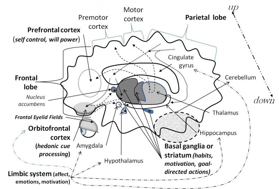
n Part 1, you were introduced to the major brain centers that are relevant to a discussion of willpower: the prefrontal cortex, the seat of will power and self control; the basal ganglia centers within the brain that process reward, store habits and goal-directed actions, and are responsible for motivation; and finally the thalamus, the transport hub connecting the basal ganglia with the higher cortical areas involved in self-control, action-planning, and behavior.
There is one more system within the brain that is highly relevant to willpower, and that is the limbic system.
You might find it helpful to once again use the brain schematic from Part 1.
The Limbic System—Emotions and Affect
Emotions are associated with brain centers in the limbic system, including the cingulate gyrus, amygdala, hypothalamus, and hippocampus. These regions of the brain process mainly negative emotions. The cingulate cortex is associated with negative emotions (fear, fright, and pain)[1]; the amygdala is linked to negative emotions (fear and anxiety)[2]; and the hippocampus is associated with the storage of emotional memories[3]. Internal cues (e.g., thoughts and memories) or external cues (someone you owe money to) that are recalled in the hippocampus[4] trigger responses in the cingulate cortex and amygdala[5], which subsequently send signals to the prefrontal cortex by way of the thalamus. The hypothalamus, influenced by signals from the amygdala, can release hormones that trigger centers elsewhere in the body[6]. Therefore, unlike the basal ganglia, limbic system elements can influence the body directly, as well as influence upper brain regions through thalamus connections. What the prefrontal cortex decides to do at any given time is strongly influenced by the inputs coming from the limbic system.
Affect
Affect is our visible physiological response to becoming aware of something pleasant, unpleasant, threatening, or motivationally significant. A sudden relaxation of the body, an increase or decrease in heart rate[7], hair standing up at the back of your neck, dilation of pupils in the eyes, or heightened attention to a stressor[8] are all examples of affective responses associated with either activation of the basal ganglia, the limbic system, or a combination of both[9].
Now, this is where everything gets interesting.
Cognitive neuroscience has shown that we do not become conscious of things in the environment immediately. There is a slight delay between the time our brain processes information about the environment and when we become aware of it. This delay is measured in milliseconds and it is critical to understanding why willpower often fails.
Priming
The activation of our brain in stages to changing environmental conditions is called priming. Priming is actually a form of nonconscious memory that happens outside of our awareness. Priming changes our ability to identify, produce, or classify an item as a result of previous experience with it or something closely related to it[10]. Priming is a critical function of the brain’s perceptual processing and classification system. It operates at a preconscious level[11] and prepares us to maximize our interaction with our surroundings.
Frontal Eyelid Fields and Parietal Lobe
Priming begins with the frontal eyelid fields and the parietal lobe. The parietal lobe is all about processing spatial information about the environment around us. The frontal eyelid field takes in the information about the environment we are moving towards and sends it to the parietal lobe, where it is processed[12]. This occurs in about 0 to 30 milliseconds. This is the first stage of priming. At this stage, the rest of the brain doesn’t know what’s going on. This function has to come first so we don’t hit anything (or fall off a cliff!).
Parietal Lobe Connections
The parietal lobe has direct connections with many other brain regions. The parietal lobe primes negative affective and emotional information associated with detected objects in the environment through connections with the front of the cingulate gyrus, the upper part of the limbic system. Parietal connections with the hippocampus of the limbic system mean that the priming effect activates memories from our past related to objects (people or things) associated with negative emotions[13]. However, we’re still not aware of them yet.
The parietal lobe also has direct connections with the front of the basal ganglia. This means the parietal lobe can prime habits and goal-directed actions[14] appropriate to detected objects (a spouse, a prospective employer, or a tray of donuts) associated with important goals or previous rewards. This occurs within about 100 milliseconds. The parietal lobe also has direct connections with and primes the orbitofrontal cortex[15].
Orbitofrontal Cortex
The orbitofrontal cortex has been associated with the limbic system because it communicates the hedonic value[16] (pleasure value, reward value) of objects detected in the local environment[17-19]. Once detected, the orbitofrontal cortex continually sends the reward/pleasure information related to those detected objects to the basal ganglia, which stimulate the production of dopamine, a key neurotransmitter associated with motivation, from the substantia nigra[20].
At the final stage, all of the information about what’s going on in the environment from the parietal lobe, the basal ganglia, and the limbic system are sent to the prefrontal cortex and premotor cortex (the action planning area). About now, we start to become aware of what is going on around us based on the full activation of the other areas of our brain.
Now, the prefrontal cortex has to make some decisions, but at this moment, what information does it have to go by?It actually has to go by awareness of the surroundings based on past experience; it has to balance the reward/pleasure aspects of the surroundings based on past experience, balance the negative emotional aspects of the environment shaped by past experience, and select from the motivated, goal-directed habits and behavioral routines coming from the basal ganglia that have proved successful in similar situations in the past.
At this point, barring any other consideration or barrier, a person will behave the same way they always have. Prior habits will run the show, complemented by minor adjustments applied by the prefrontal cortex. The prefrontal cortex’s exercise of self-control and willpower will be to make adjustments, select the appropriate actions based on the situation, and inhibit those behaviors and habits that don’t quite fit.
In situations like this, there are rarely failures of willpower as the self-control demands aren’t that daunting. The major lifting is done by habits.
It’s another story, however, if a person decides that it’s time to abruptly change behavior.
Suppose that a person decides to lose weight or adopt a new diet. The first few days of the diet generally go well. After several days, however, the person starts to crave the foods he or she has given up. Willpower begins to crack. As days pass, the crack grows wider. Any stressor, no matter how small, weakens it further. Pretty soon, the person is paying lip service to the diet, seeing no progress, and still questioning why he or she couldn’t stick to it. Why did willpower fail?
Continued in Part 3.
[expand title=”References (click to expand)”]
- Shackman, A. J., Salomons, T. V., Slagter, H. A., Fox, A. S., Winter, J. J., & Davidson, R. J. The integration of negative affect, pain and cognitive control in the cingulate cortex.Nature Reviews Neuroscience. 2011; 12(3), 154-167.
- Lang, P. J., & Bradley, M. M. Emotion and the motivational brain.Biological Psychology. 2010; 84(3), 437-450.
- Murty, V. P., Ritchey, M., Adcock, R. A., &LaBar, K. S. fMRI studies of successful emotional memory encoding: a quantitative meta-analysis. Neuropsychologia. 2010; 48(12), 3459-3469.
- Rossato, J. I., Bevilaqua, L. R., Izquierdo, I., Medina, J. H., &Cammarota, M. Dopamine controls persistence of long-term memory storage. Science. 2009; 325(5943), 1017-1020.
- Roozendaal, B., McEwen, B. S., & Chattarji, S. Stress, memory and the amygdala. Nature Reviews Neuroscience. 2009; 10(6), 423-433.
- Herman, J. P., Ostrander, M. M., Mueller, N. K., &Figueiredo, H. Limbic system mechanisms of stress regulation: hypothalamo-pituitary-adrenocortical axis. Progress in Neuro-Psychopharmacology and Biological Psychiatry. 2005; 29(8), 1201-1213.
- Kirsch, D. L. The physiological response of affect. 1999, May 24; https://vismod.media.mit.edu/tech-reports/TR-495/node7.html
- Lang, P. J. The emotion probe: studies of motivation and attention. American Psychologist. 1995; 50(5), 372.
- Chiew, K. S., & Braver, T. S. Positive affect versus reward: emotional and motivational influences on cognitive control.Frontiers in Psychology. 2011; 2, 279.
- Schacter, D. L., Dobbins, I. G., &Schnyer, D. M. Specificity of priming: A cognitive neuroscience perspective. Nature Reviews Neuroscience. 2005; 5(11), 853-862.
- Tulving, E., &Schacter, D. L. Priming and human memory systems.Science. 1990; 247(4940), 301-306.
- Lane, A. R., Smith, D. T., Schenk, T., & Ellison, A. The involvement of posterior parietal cortex and frontal eye fields in spatially primed visual search. Brain Stimulation. 2012; 5(1), 11-17.
- Wagner, A. D., Shannon, B. J., Kahn, I., & Buckner, R. L. Parietal lobe contributions to episodic memory retrieval.Trends in Cognitive Sciences. 2005; 9(9), 445-453.
- Tucker, M., & Ellis, R. Action priming by briefly presented objects.ActaPsychologica. 2004; 116(2), 185-203.
- Jarbo, K., &Verstynen, T. D. Converging structural and functional connectivity of orbitofrontal, dorsolateral prefrontal, and posterior parietal cortex in the human striatum. The Journal of Neuroscience. 2015; 35(9), 3865-3878.
- Elliott, R., Agnew, Z., & Deakin, J. F. W. Hedonic and informational functions of the human orbitofrontal cortex.Cerebral Cortex. 2010; 20(1), 198-204.
- Kringelbach, M. L. The human orbitofrontal cortex: linking reward to hedonic experience. Nature Reviews Neuroscience. 2005; 6(9), 691-702.
- Beer, J. S., John, O. P., Scabini, D., & Knight, R. T. Orbitofrontal cortex and social behavior: integrating self-monitoring and emotion-cognition interactions. Cognitive Neuroscience, Journal of. 2006; 18(6), 871-879.
- Rolls, E. T. The orbitofrontal cortex and reward.Cerebral Cortex. 2000; 10(3), 284-294.
- Ponzi, A. Dynamical model of salience gated working memory, action selection and reinforcement based on basal ganglia and dopamine feedback. Neural Networks. 2008; 21(2/3), 322-330.
[/expand]


